INTRODUCTION
Oxidative stress is recognized as one of the key drivers of skin ageing as it is linked to DNA damage, lipid and protein oxidation, and also to the accumulation of advanced glycation end products (AGEs) (1). AGEs are formed during glycation reactions where reducing sugars react with free amino groups of proteins. In the skin, this reaction involves the cross-linking of sugars with dermal extracellular matrix proteins like collagen, impairing their structure and elastic properties, contributing to skin ageing. Furthermore, AGEs promote the generation of reactive oxygen species (ROS) and pro-inflammatory cytokines, exacerbating inflammatory responses in the skin ( 2). This in turn triggers the accumulation of melanin and lipofuscin, contributing to the yellowish discolouration of aged skin and to the formation of age spots(3). NRF2, which stands for Nuclear factor erythroid 2-related factor 2, is a transcription factor that plays a critical role in the body’s antioxidant response regulating the expression of numerous genes involved in cellular defence against oxidative stress and inflammation (4). NRF2 activation has been shown to have protective effects against oxidative damage to the skin helping reduce stress and the formation of age spots. In this scenario, we selected an extract derived from the species Jasminum sambac for its interesting antioxidant properties. In particular, as reported by Ceccacci et al in 2022 (5), we developed a hydroethanolic extract derived from Jasminum sambac plant cell culture (JsEx) and characterized it for its chemical profile, revealing the presence of active compounds like polyphenols or lignans with known antioxidant and anti-inflammatory properties. We revealed the ability of the extract to enhance the NRF2 pathway and as consequence to reduce ROS production in keratinocytes and to inhibit the inflammatory cascade in LPS-stimulated macrophages. We demonstrated a decrease in AGE formation in glyoxal treated fibroblasts and in methylgyoxal stressed skin explants. In addition to this, JsEx also showed an extracellular matrix boosting effect, increasing the production of collagen type I, which is highly sensitive to glycation (6) and whose levels are significantly reduced by oxidative stress (7).
As ethnic origin greatly influences skin biology and the way our skin reacts to pigmentation disorders (8), this study aimed to clinically assess the biological power of Jasminum sambac cell extract to counteract skin ageing and skin hyperpigmentation in two different panels of healthy volunteers, Asian and Caucasian. We finally demonstrated that, independently of skin tone, Jasminum sambac cell extract was able to improve skin complexion and regulate pigmentation.
MATERIALS AND METHODS
Melanin Content assay
Melanocytes (B16F1) were seeded in 96-well plates at a density of 3 x 10³ and grown for 20 hours. Then, the cells were treated with JsEx in presence of IBMX 50 mM for 72 hours. At the end of the treatment, the cells were washed with PBS, and lysed in 50 ml of NaOH 1M at 70°C for 20 min. The absorbance values were measured using the instrument Victor Nivo (PerkinElmer Inc., Singapore). The bars represent the standard deviations, and the asterisks indicate the p-value according to Student’s t test (* p < 0.05, ** p < 0.01, *** p < 0.001).
Clinical studies
The cosmetic effects of the Jasminum sambac cell culture extract were evaluated through a mono-centric, double blind clinical study performed by an independent lab in a panel of 22 healthy volunteers, aged from 35 to 65 years old, 50% Caucasian and 50% Asian. Specific inclusion criteria were age from 35 and 65 years, female, with a phototype (Fitzpatrick): I, II, III or IV, with dull complexion and dark age spots on face.
The subjects were randomly assigned to apply a formulation containing 0.5% w/w Jasminum sambac cell extract (JsEx) previously dissolved in glycerol or a placebo formulation on each half face twice daily for 42 days.This JsEX concentration in the formulation mirrors that used in the in vitro assay as reported by Ceccacci et al in 2022 (5). The comparative assessment of the cosmetic efficacy was conducted through a combination of instrumental measurements and clinical evaluations at three time points: baseline (D0), 28 days (D28) and 42 days (D42) after the initiation of product use. Skin colour was determined using the Colorimeter CL 400® (Courage & Khazaka), while photographs of the skin were taken with the Antera 3D® camera (Miravex) to evaluate dark spots and analyze the parameters “melanin” and “skin texture” using dedicated software. Clinical examinations and grading were carried out through live objective evaluation by a blinded dermatologist, focusing on complexion radiance, homogeneity and pigmentary spots. Instrumental measurements were performed by technicians under the dermatologist’s supervision. All the examinations following established criteria were performed on each half of the face at D0, D28, and D42.
The test was conducted according to the principles of the Helsinki Declaration (1964) and subsequent revisions endorsed by the European Community and according to Colipa Guidelines for the evaluation of the efficiency of cosmetic products.
Statistical analysis
The following data were calculated: the average and the standard deviation at each experimental time, the variations D28 vs D0/T0 and D42 vs D0/T0, the variations in percentage D28 vs D0/T0 and D42 vs D0/T0, the distribution of difference (Ti-T0) by a Shapiro-Wilk test, at risk of 5%. The significance was calculated according to p-value of bilateral Student t test in pairs, in case of normality of the distribution or the Wilcoxon test in the adverse case. The statistical significance in variations of D28 and D42 between placebo group and verum group were also calculated based on Wilcoxon test. The variations are judged statistically significant if p<0.05.
RESULTS
Melanin content in melanocytes
A cell culture of melanocytes stimulated with IBMX to boost melanin production was treated with JsEx to determine its ability to regulate melanin synthesis. As shown in figure 1, JsEx used at 0.0006% w/v reduced the release of melanin by 28% at level comparable to kojic acid used as positive control.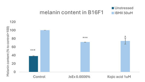
Figure 1. Melanin content in B16F1 treated with JsEx or Kojic acid together with IBMX for 72 hours. The values reported in the graph represent the percentage with respect to IBMX stimulated cells set as 100%. Each value represents the average of three independent experiments each of them being performed in triplicate. *** indicates pvalue <0.001 and * means pvalue <0.05.
Colorimetry determination
The facial skin colour was determined using the Colorimeter CL400® (Courage & Khazaga) to evaluate the Individual Typology Angle ITA° and the b*. The ITA°, according to the formula ITA°= [Arc Tangent ((L* – 50) / b*)] 180 / 3.14159 where b* is the yellow-blue axis, is a measure of an individual’s skin colour based on the angle of their skin undertone relative to a standard colour space. Both after 28 and 42 consecutive days of product use (D28 and D42), no statistically significant modification in the b* and ITA° was observed in the group of people treated with placebo whereas in volunteers treated with the formula containing Jasminum extract, b* decreased by 6.88% after 28 days and by 8.75% after 42 days. Also, in the group treated with the formula containing the extract, the ITA° significantly improved, increasing by 4.07% after 28 days and 6.05% after 42 days with respect to T0. In particular the difference between the variation of the b* and ITA° values on the group treated with the Jasminum sample and the group treated with the placebo was statistically significant in favour of the first group at both the measured time point D28 and D42.
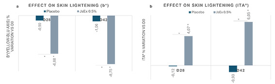
Figure 2 Effect on skin lightening evaluated by Colorimeter CL400. 2a the percentage difference in b* between 28 or 42 days of treatment and time 0. 2b: the percentage difference in ITA° between 28 or 42 days of treatment and time 0.
Evaluation of dark spots
In order to evaluate the effect of the formulation containing JsEx on dark spots, photographs of the skin taken with the camera Antera 3D® (Miravex) were analysed with dedicated software that measures the average concentration and uniformity of melanin and haemoglobin, including their hyper- and hypo- concentrations, as well as skin texture (9).
Comparing the photographs obtained before (D0/T0) and after 28 and 42 consecutive days of product use (D28 and D42), a statistically significant reduction of the dark spots by 4.49 after 28 days and 6.66 after 42 days for a lighter and more luminous skin were noted in volunteers treated with JsEx. Moreover, a statistically significant reduction of skin roughness was measured in volunteers treated with JsEx resulting in more homogeneous and smoother complexion. The variation between people treated with JsEx and people treated with placebo formula was also statistically significant.
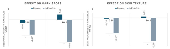
Figure 3. Clinical evaluation of pictures taken with the camera Antera 3D® (Miravex). 3a: effect on dark spots expressed as melanin content % variation vs D0 at D28 and D42. 3b: Effect on Skin texture expressed as skin roughness percentage difference versus D0 at D28 and D42.
After both 28 and 42 consecutive days of product use, the sample containing JsEx induced a statistically significant decrease in the dark spots and skin roughness versus time 0. * means pvalue <0.5
Objective evaluation by the dermatologist
The third analysis performed on volunteers involved in the double-blind study was the objective evaluation by the dermatologist of the following parameters: complexion radiance, complexion homogeneity and pigmentary spots.
The table 1 shows that the sample treated with the Jasminum formula statistically significantly improved parameters of complexion radiance, complexion homogeneity and pigmentary spots after both 28 and 42 consecutive days of product use. The difference between the variation in all the recorded parameters on the group treated with the active formula with respect to the sample treated with the placebo was statistically significant.
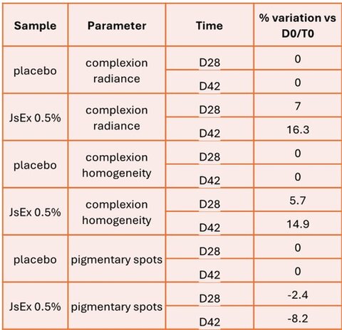
Table 1. Clinical score assigned.
Figure 4 presents some representative HD photos from two groups of volunteers, where the reduction in pigmentary spots was evident.

Figure 4. Representative photographs from Asian or Caucasian volunteers treated with formula containing JsEx. The arrows indicate visible spots appearance over time.
DISCUSSION AND CONCLUSIONS
Jasminum sambac, an Oleaceae family member native to India, is renowned for its historical use and significant role in Ayurvedic medicine. This study aimed to isolate beneficial molecules from the whole plant using suspension cells, following Ceccacci et al. (2022) (5). Plant cell cultures are used in cosmetics as a sustainable and eco-friendly system to derive secondary plant metabolites of skin care interest (10;11). These cultures can be optimized to enhance the yield and potency of desired compounds, offering a consistent and controlled source of bioactive ingredients.
Jasminum sambac suspension cells were used to generate a hydroethanolic extract very rich in beneficial compounds, with the ability to regulate pathways like NRF2 and, as a consequence, to reduce reactive oxygen species (ROS) and counteract the production of Advanced glycation end products (AGEs). Given these very promising results (5) we investigated the application of the extract as a multi-faceted approach to address hyperpigmentation and uneven skin tone. In vitro analysis demonstrated that the JsEx reduces melanin synthesis. Based on these findings, we further evaluated its effects through clinical testing.
Skin colour results from the action of multiple biological pathways that primarily control melanin synthesis and its distribution. Ageing affects nearly every aspect of cutaneous biology, including melanin accumulation and pigmentation which often results in uneven skin colouring (or hyperpigmentation). Factors such as excessive oxidative stress can lead to unbalanced formation of advanced glycation end products (AGEs), yellowish substances formed in the late stage of glycation affect all levels of the skin, causing inflammation, ageing, pigmentation disorders and other issues and it is well known that accumulation of AGEs is associated with the development or worsening of many degenerative processes.
Cutaneous manifestation of ageing is strongly influenced by ethnic origin as different skin types have structural and functional differences. Considering the influence of both intrinsic and extrinsic factors on cutaneous effects and their variation based on ethnic origin (12), we conducted a clinical trial under dermatological control involving a diverse group of volunteers from two different ethnic backgrounds, encompassing both Caucasian and Asian participants with varied skin tones and conditions. This ensured the broad applicability and robustness of our findings, despite a modest sample size. The overall conclusion of clinical studies revealed that the formula containing the Jasminum extract statistically decreases the b* value, increases the ITA° value, decreases melanin and improves skin texture, complexion radiance and homogeneity and reduces pigmentary spots visibility. These results, strongly supported by previously reported in vitro test, highlight the ability of the Jasminum extract to regulate pigmentary reactions in the skin.
In conclusion Jasminum sambac cell culture extract is a biotechnology based active ingredient able to boost skin antioxidant defences and prevent the glycation process in the skin. This synergistic effect ensures a powerful effect on skin complexion, fighting skin pigmentation disorders and skin roughness.
References and notes
- Rinnerthaler M, Bischof J, Streubel MK, Trost A, Richter K. Oxidative Stress in Aging Human Skin. Biomolecules. 2015; 5(2):545-589. Available from https://www.mdpi.com/2218-273X/5/2/545
- Twarda-Clapa A, Olczak A, Białkowska AM, Koziołkiewicz M. Advanced Glycation End-Products (AGEs): Formation, Chemistry, Classification, Receptors, and Diseases Related to AGEs. Cells. 2022; 11(8):1312. Available from https://www.mdpi.com/2073-4409/11/8/1312
- Skoczyńska A, Budzisz E, Trznadel-Grodzka E, Rotsztejn H. Melanin and lipofuscin as hallmarks of skin aging. Advances in Dermatology and Allergology/Postępy Dermatologii i Alergologii. 2017;34(2):97-103. Available from https://doi.org/10.5114/ada.2017.67070
- Frantz MC, Rozot R, Marrot L. NRF2 in dermo-cosmetic: From scientific knowledge to skin care products. Biofactors. 2023;49(1):32-61. Available from https://doi.org/10.1002/biof.1907
- Ceccacci S, Lucia AD, Tortora A, Colantuono A, Carotenuto G, Tito A, Monti MC. Jasminum sambac Cell Extract as Antioxidant Booster against Skin Aging. Antioxidants. 2022; 11(12):2409. Available from https://doi.org/10.3390/antiox11122409
- Hudson DM, Archer M, King KB, Eyre DR. Glycation of type I collagen selectively targets the same helical domain lysine sites as lysyl oxidase-mediated cross-linking. J Biol Chem. 2018;293(40):15620-15627. Available from 10.1074/jbc.RA118.004829
- Tu Y, Quan T. Oxidative Stress and Human Skin Connective Tissue Aging. Cosmetics. 2016; 3(3):28. Available from https://doi.org/10.3390/cosmetics3030028
- Naik PP, Farrukh SN. Influence of Ethnicities and Skin Color Variations in Different Populations: A Review. Skin Pharmacol Physiol. 2022;35(2):65-76. Available from https://doi.org/10.1159/000518826
- Messaraa C, Metois A, Walsh M, et al. Wrinkle and roughness measurement by the Antera 3D and its application for evaluation of cosmetic products. Skin Res Technol. 2018;24(3):359-366. Available from doi:10.1111/srt.12436. https://onlinelibrary.wiley.com/doi/10.1111/srt.12436
- Tito A., Barbulova A., Zappelli C. et al. The Growth Differentiation Factor 11 is Involved in Skin Fibroblast Ageing and is Induced by a Preparation of Peptides and Sugars Derived from Plant Cell Cultures. Mol Biotechnol 61, 209–220 (2019). Available from https://link.springer.com/article/10.1007/s12033-019-00154-w
- Tito A, Bimonte M, Carola A, et al. An oil-soluble extract of Rubus idaeus cells enhances hydration and water homeostasis in skin cells. Int J Cosmet Sci. Available from https://pubmed.ncbi.nlm.nih.gov/25940647/
- Vashi NA, de Castro Maymone MB, Kundu RV. Aging Differences in Ethnic Skin. J Clin Aesthet Dermatol. 2016 Jan;9(1):31-8. PMID: 26962390; Available from https://www.ncbi.nlm.nih.gov/pmc/articles/PMC4756870/
Unleashing Nature’s Potential: The Future of Sustainable Beauty...

Cyclization as a tool for reinvention

Cosmetics for me – Personalized products: Opportunities and Limi...
Navigating the Balance – Personalized Skincare and Consumer Perc...

Is Microbiome Certification even Necessary?

Microbiome-friendly – The How and Why of this claim
ISNTREE – Yam Root Vegan Milk Toner