LAURIE VERZEAUX*, LAETITIA MARCHAND, ELODIE AYMARD, HÉLÈNE MUCHICO, BRIGITTE CLOSS
*Corresponding author
SILAB, Brive, France
Introduction
The world’s population is aging at an accelerated rate, explaining the ever-increasing demand from consumers for products and services that enable them to age in good health. This demand is stimulated by scientific progress in preventive medicine and cutting-edge technologies that enhance our understanding of biological aging (1). The science of longevity is of outmost importance for researchers and in parallel it opens the path to new perspectives for the concept of timeless beauty and “anti-aging” products. Research on longevity promises a caring and rational support to female consumers weary of the ideals of a youthful appearance. Research has shown that the aging process results from the slowing of biological functions (2). Mitochondrial functions in the epidermis are disrupted, leading to the progressive installation of oxidative stress, a loss of energy production and ultimately a reduced rate of epidermal renewal (3). In the dermis, harmful biological phenomena appear, such as the glycation of proteins or the accumulation of senescent cells. The quality of the extracellular matrix deteriorates, leading to slackening of the skin and the appearance of wrinkles.
Scientific investigations conducted to elucidate the mysteries of longevity have revealed mechanisms and markers that can limit the decline of cellular functions (4,5). The family of sirtuins has therefore been the focus of research on longevity for more than 20 years because they have been defined as promoting life expectancy (6) . Historically discovered in a yeast, sirtuin 1 is the first to have demonstrated a capacity to prolong the lifetime of an organism. Since then, the biology of sirtuins has continued to raise interest of scientists and has resulted in a large number of publications on the relationship between the other members of this family and longevity (7). The longevity promoting activity of sirtuins depends on the availability of the coenzyme NAD+, required for correct mitochondrial functioning and energy production, and of AMPK in its active phosphorylated form, an energy sensor that protects cell metabolism (8). Members of the sirtuins family stand out by their varied cellular localizations and their transversal biological activities. These properties enable their beneficial actions on many mechanisms related to aging (5,9). Sirtuins 3 and 6 therefore positively affect mitochondrial activity by reducing oxidative stress that is harmful to correct cell functioning (10, 11, 12). Sirtuins 1 and 7 have been shown to limit glycation and delay the onset of cell senescence, two mechanisms essential for preserving the quality of the dermis (7,13). Based on these properties, sirtuins are currently considered as strategic targets for increasing cell longevity. In this context, the aim of this study was to investigate the mechanism of action of a natural cosmetic active ingredient (INCI: Myrtus communis leaf extract) on sirtuins and their biological consequences at the level of the epidermis and the dermis.
Material and methods
1. Effect of the Myrtus communis leaf extract on epidermal functioning
2D in vitro model
Studies were conducted on keratinocytes from old donors (> 60 years of age), seeded and incubated at 37°C in an atmosphere containing 5% CO2. Keratinocytes were then treated or not with the Myrtus communis leaf extract.
3D in vitro model
Reconstructed epidermis (SILABSKIN® RE) were prepared from young human keratinocytes (P1) and from keratinocytes aged by successive replications (P9). Human keratinocytes were seeded in inserts and incubated at 37°C in an atmosphere containing 5% CO2. After 14 days of culture, SILABSKIN® RE were treated or not with the Myrtus communis leaf extract during 48h.
Study of sirtuins co-activators
The level of AMPK phosphorylation (AMPK-P) was quantified in old keratinocytes after 24h of treatment with the natural active ingredient by capillary Western blot and analyzed using Compass software v4.1.0 (ProteinSimple). The ratio of the quantity of AMPK-P over the total quantity of AMPK was calculated and is expressed as arbitrary units (AU).
Intracellular NAD+ production was also quantified in keratinocytes after 24h of treatment with the natural active ingredient by using the NAD/NADH-GloTM Assay Kit (Promega). The quantity of intracellular NAD+ was determined using a standard range of pure NAD+ and is expressed as nanomolar (nM).
Study of sirtuins syntheses
SILABSKIN® RE were recovered, fixed, dehydrated, embedded in paraffin, and sections were prepared with a microtome. The syntheses of SIRT3 and SIRT6 were determined by immunohistofluorescence. Quantitative analysis of the images was conducted using an image analysis script developed in-house with Python language. The results are expressed in arbitrary units (AU).
Mitochondrial dynamics
Probes were used to visualize mitochondrial dynamics. MitoTrackerTM Green FM was used to label mitochondria, Hoechst for cell nucleus and CellMaskTM Orange for plasma membrane. Visualization was done with an LSM700 confocal microscope (Zeiss), coupled to a Zen Black image Analysis system (Zeiss). Staining the plasma membrane and the nucleus was used for the automatic detection of cells during quantification. The appearance ratio parameter of the mitochondrial network was quantified in each cell with the MITOSHAPE® program, implemented using Matlab® software, release R2012b (MathWorks) and Fiji image analysis software release 1.51g (14). The appearance ratio is the ratio of mitochondrion length/mitochondrion width. The appearance ratio decreases with increasing fragmentation of the mitochondrial network.
Mitophagy
This study was conducted by immunocytofluorescence on old human keratinocytes subjected for 24h to a free radical stress by treatment with CCCP (Carbonyl cyanide m-chlorophenylhydrazone) at 1µM (old stressed keratinocytes). CCCP is a powerful agent that depolarizes the mitochondrial membrane and is known to induce mitophagy. These cells were then incubated in the presence or absence of the natural active ingredient and incubated at 37°C in an atmosphere containing 5% CO2. Mitophagy was then quantified thanks to an immunocytofluorescence conducted with anti-GRP75 and anti-LCB3 antibodies. Visualization of mitochondria (green) and autophagosomes (red) was done with an LSM700 confocal microscope (Zeiss), coupled to a Zen Black image analysis system (Zeiss). When the two immunolabelings are superimposed, mitophagosomes appear yellow. The percentage of mitophagosomes was quantified using a classical digital analysisprogram implemented in Fiji software, release 1.51g to study colocalization between masks relative to mitochondria and autophagosomes, generated by an automatic threshold. The percentage of mitophagosomes is the number of autophagosomes containing a mitochondrion with respect to the total number of autophagosomes in cells. Mitophagy results in an increase in the number of mitophagosomes, expressed as percentage.
2. Effect of the Myrtus communis leaf extract on the dermal matrix
2D in vitro model
Human fibroblasts were seeded and incubated at 37°C in an atmosphere containing 5% CO2 for 3 days. Then, cells were daily treated with a solution of hydrogen peroxide (H2O2) for 4 days in the presence or absence of the Myrtus communis leaf extract.
Study of sirtuins co-activators
The level of AMPK-P and of NAD+ were assayed as previously described.
Study of sirtuins syntheses
The syntheses of SIRT1 and SIRT7 were quantified by capillary Western blot as previously described. The rate of proteins of interest is expressed in AU and is the ratio of proteins of interest/total proteins.
Cell senescence
A cell staining kit (Abcam) with an Axio-Observer Z1 microscope (Zeiss) coupled to an AxioCam MRc camera (Zeiss) Zen Blue analysis software (Zeiss) were used to visualize SA-β-galactosidase activity. DAPI labeling was used to obtain cell counts and the ratio of the number of positive cells showing β-galactosidase staining near the nucleus. Quantitative analysis of the images was conducted using an image analysis script developed in-house with Python language. The results are expressed as percentage of SA-β-galactosidase-positive cells.
3. Cosmetic benefits of the Myrtus communis leaf extract
This study was conducted in accordance with Good Clinical Practice (GCP). Volunteers were selected according to the following criteria : between 50 and 70 years of age, the presence of crow’s feet wrinkles (stage between 2 and 4 according to a scoring scale from 1: absence of wrinkles to 6: presence of many wrinkles), a dull complexion (stage less than 3 according to a scoring scale from 1: very dull complexion to 5: radiant complexion) and an elevated glycation rate (cutaneous autofluorescence ≥ 1.8 AU). The Myrtus communis leaf extract was formulated at 2% in an emulsion and vehicle controlled studies were conducted. The Caucasian panel was composed of two groups, a vehicle controlled group with 20 volunteers, mean age 61 ± 7 years, and a group evaluating the natural active ingredient with 21 volunteers, mean age 58 ± 6 years. Volunteers applied the emulsion to the entire face twice daily for 14 days. The skin microrelief was investigated by fringe projection (AEVA-HE²) and the complexion radiance was evaluated thanks to visual scoring by trained evaluators. The Asian panel was composed of 34 volunteers, mean age 60 ± 5 years. Volunteers applied the emulsion to each half of the face twice daily for 56 days. Wrinkles were evaluated thanks to clinical scoring by a dermatologist and skin elasticity was investigated by subjective evaluation.
4. Statistical analysis
The results between the different measurement times in the same group were compared with Student’s t test for paired data when the data followed a normal law (Shapiro-Wilk test of normal distribution more than 5%) or if not with the non-parametric Wilcoxon signed ranks test. The results obtained between the two treatments were compared with Student’s t test for independent data when the data followed a normal law or if not with the non-parametric Mann-Whitney test. In each case, the test was one-tailed with a risk threshold set at 5%. This statistical processing was done on Excel with the Real Statistics library (release 7.2, Copyright (2013-2020), Charles Zaiontz).
Results
1. Effect of the Myrtus communis leaf extract on epidermal functioning
The longevity-promoting activity of sirtuins depends on the availability of their coactivators AMPK in its activated phosphorylated form and NAD+. Tested at 0.10% on old keratinocytes, the Myrtus communis leaf extract significantly increases the AMPK phosphorylation and the NAD+ synthesis by 55% (figures 1A and B).
Moreover, the SIRT3 and SIRT6 syntheses are significantly reduced during aging (data not shown). Tested at 0.10% on aged SILABSKIN® RE, the Myrtus communis leaf extract significantly restores the syntheses of these two sirtuins by 103% and 71%, respectively (figures 1C and D).
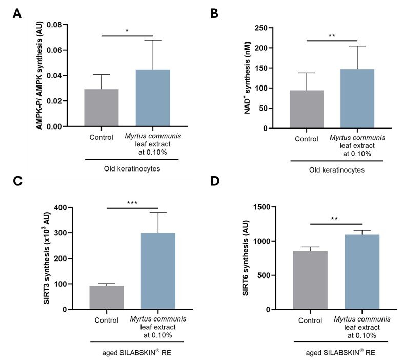
Figure 1. Effect of the Myrtus communis leaf extract on the phosphorylation of AMPK (A) and on NAD+ synthesis (B) by old keratinocytes. Effect of the Myrtus communis leaf extract on the SIRT3 (C) and SIRT6 (D) syntheses by aged SILABSKIN® RE. Result(s) with *: P < 0.05 — **: P < 0.01 — ***: P < 0.001.
In the case of moderate cell damage, the dynamic properties of mitochondria become involved. In fact, these organelles have the capacity to constantly remodel, thus maintaining a functional mitochondrial population (15). This remodeling involves two mechanisms. Fusion pools the contents of moderately damaged mitochondria with healthy mitochondria to maintain a uniform and functional mitochondrial population. Fission is a sequestering phenomenon corresponding to the fragmentation of an extensively damaged mitochondrion to form a healthy mitochondrion and a damaged mitochondrion that will be eliminated by mitophagy. The appearance ratio is the ratio of mitochondrion length/mitochondrion width. The appearance ratio decreases with increasing fragmentation of the mitochondrial network. With aging, the morphology of the mitochondrial network is modified with a significant reduction in old human keratinocytes. Tested at 0.10% on old keratinocytes, the Myrtus communis leaf extract significantly restores the appearance ratio by 103% (figure 2A).
In the case of substantial damage that could be fatal to cell survival, mitochondria are selectively eliminated by mitophagy. This biological process is a form of autophagy that specifically targets damaged mitochondria by autophagosomes. This leads to the formation of mitophagosomes and, to the elimination of defective mitochondria. The formation of mitophagosomes was analyzed by determining the colocalization of the markers GRP75 (mitochondria) and LC3B (autophagosomes), direct proof that mitochondria are being eliminated by mitophagy. Tested at 0.050% on old keratinocytes subjected to free radical stress, the Myrtus communis leaf extract significantly increases the formation of mitophagosomes by 39%, therefore improving mitochondrial activity by eliminating defective mitochondria (figure 2B).
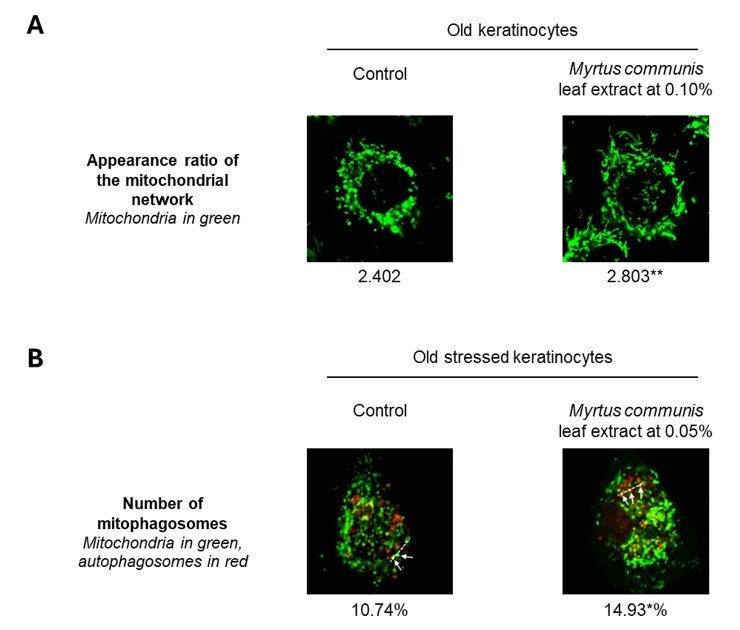
Figure 2. Effect of the Myrtus communis leaf extract on the appearance ratio of the mitochondrial network of old human keratinocytes (A) and on the mitophagy in old, stressed keratinocytes (B). Result(s) with *: P < 0.05 —
**: P < 0.01.
The production of intracellular ATP by mitochondria was then assayed by luminescence in old keratinocytes. Results reveal that tested at 0.10%, the Myrtus communis leaf extract significantly restores the ATP production by 50% in old keratinocytes (data not shown). This natural active ingredient therefore reactivates energy production required for correct cell functioning: the proliferation of old keratinocytes is restored, as well as the syntheses of differentiation and cohesion markers (data not shown).
2. Effect of the Myrtus communis leaf extract on the dermal matrix
The AMPK-P and NAD+ levels were assessed in fibroblasts subjected to an H2O2 stress. The Myrtus communis leaf extract at 0.10% significantly increases the phosphorylation of AMPK and the synthesis of NAD+ by 42% and 35%, respectively (figures 3A and B). Moreover, tested at 0.10% on the same model, this natural active ingredient significantly increases the syntheses of SIRT1 and SIRT7 by 95% and 115% respectively (figures 3C and D).
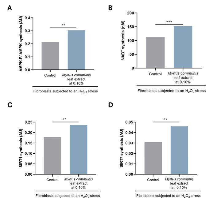
Figure 3. Effect of the Myrtus communis leaf extract on the phosphorylation of AMPK (A), NAD+ synthesis (B) by fibroblasts subjected to an H2O2 stress. Effect of the Myrtus communis leaf extract on the syntheses of SIRT1 (C) and SIRT7 (D) by fibroblasts subjected to an H2O2 stress. Result(s) with **: P < 0.01 — ***: P < 0.001.
Then, its capacity to limit the onset of cell senescence was investigated. When the Myrtus communis leaf extract at 0.10% was applied at the same time as the senescence-inducing stress, it significantly limits the number of SA-β-galactosidase-positive cells by 50% (figure 4A). At the level of the dermal matrix, this natural active ingredient preserves the collagen I and elastin networks by 39% and 94%, respectively (figure 4B). Moreover, the skin glycation is limited and the quality of the dermal matrix is significantly improved in vivo (data not shown).
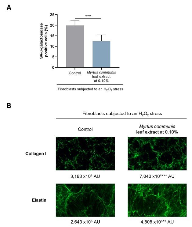
Figure 4. Effect of the Myrtus communis leaf extract on the onset of senescence of fibroblasts subjected to an H2O2 stress (A). Effect of the Myrtus communis leaf extract on the collagen I and elastin networks of fibroblasts subjected to an H2O2 stress (B). Result(s) with **: P < 0.01 — ***: P < 0.001.
3. Cosmetic benefits of the Myrtus communis leaf extract
Formulated at 2% in an emulsion, the in vivo efficacy of the Myrtus communis leaf extract was investigated in vehicle controlled studies conducted in Caucasian and Asian volunteers. After 14 days of twice daily application by Caucasian volunteers and compared to the placebo, the Myrtus communis leaf extract significantly reduces the parameter of density of surface irregularities reflecting the smoothing of microrelief by 15.1% under the eyes and 12.7% on the cheeks (figure 5A). Moreover, compared to the placebo, this natural active ingredient significantly improves the parameters characteristic of complexion radiance in Caucasian volunteers (figure 5B). These results are also observed in Asian volunteers. Tested on this panel of volunteers, Myrtus communis leaf extract also significantly reduces the wrinkles under the eyes by 6.2% (figure 5C) and improves skin elasticity by 13.3% after 56 days of treatment (figure 5D).
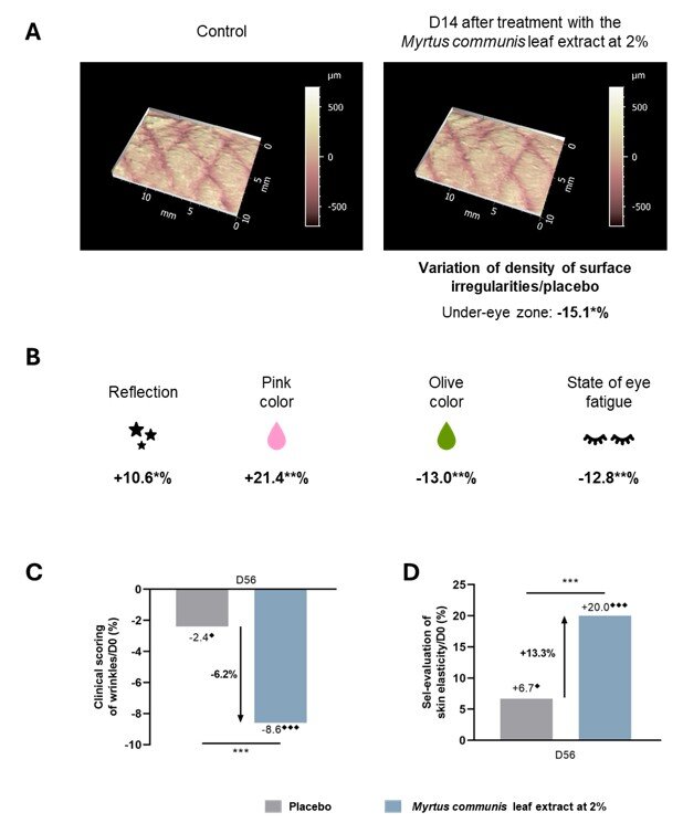
Figure 5. Effect of the Myrtus communis leaf extract on the skin microrelief (A) and the complexion radiance (B) of Caucasian skin after 14 days of treatment. Effect of the Myrtus communis leaf extract on wrinkles (C) and skin elasticity (D) of Asian skin after 56 days of treatment. Result(s)/placebo with *: P < 0.05 — **: P < 0.01 —
***: P < 0.001. Result(s)/D0 with ⬧: P < 0.05 — ⬧⬧⬧: P < 0.001.
Conclusion
Hence, by increasing the syntheses of sirtuins, and their coactivators, the Myrtus communis leaf extract activates the biological pathways of the epidermis and the dermis promoting skin functionalities for longevity. Energy production is enhanced and oxidative stress is limited in the epidermis by boosting mitochondrial function. Moreover, the dermal matrix is preserved in the dermis. At the level of the skin, microrelief is smoothed and complexion radiance is boosted. Moreover, wrinkles are reduced, and skin elasticity is improved. As a genuine promoter of cutaneous cell longevity, this natural active ingredient is recommended in all anti-aging face and body care products. This study thus focusses on the longevity and the role of sirtuins, it could also be interesting to investigate other biological markers and pathways involved in aging. It could be related to one or various of the
12 hallmarks of aging, interesting to target to counteract skin aging and promote skin longevity.

Precision Fermentation Meets AI: Transforming Personal Care Ingredient...

Exploring Hybrid Beauty: from the origins to the latest trends and reg...
Skinimalism, Hybrid Beauty and What Consumers Really Want
Soothing and Protective effects of a mixture of Saccharide Isomeratem...

The complexity of sunscreens

Patents for Chemists: A Practical Guide to Innovation Protection
EUDR – Europe’s Dumbest Regulation
The Green Transformation: Strategies to Achieve Net Zero in the Househ...